Iron Overload in Sickle Cell Disease
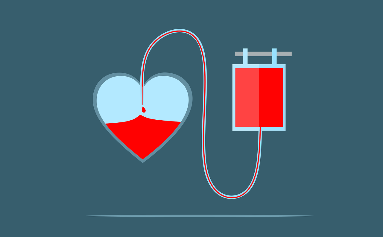
On World Sickle Cell Day, we’re encouraging people to take action by donating blood. People with sickle cell disorder may have a lifelong reliance on blood transfusions, and they are an important part of treatment for the condition.
Over time, though, people with sickle cell disorder may experience a few complications, such as iron overload. This article looks at how it arises, and the treatments available.
Why Blood transfusions?
Sickle cell disease (SCD) is the most common cause of stroke in early childhood and 24% of sickle cell patients have a stroke by the age of 45. [1] There is an increased risk for stroke in sickle cell patients because sickled red blood cells have a tendency to clot together, blocking blood vessels and causing a stroke when moving to the brain.
For patients deemed at risk, regular blood transfusions reduce the likelihood of clotting and therefore stroke.
Blood transfusions are also used on occasions to help treat other complications of SCD such as acute chest syndrome, acute anaemia, or acute priapism[2].
Our Sickle Cell Society Give Blood Send Love programme reaches out to get more people to give blood. We focus on black heritage communities to: raise awareness of the need for ethnically matched blood to treat people with sickle cell; increase levels of confidence in giving blood and recruit new donors to the blood donation register.
Ethnically matched blood is required as it is less likely to be rejected by people having frequent blood transfusions, and some blood types, such as the Ro subtype, are more commonly found in people of African and Caribbean heritage than people with white heritage.
How do Blood transfusions cause Iron Overload?
Blood transfusions are an important part of sickle cell care, and we strive to encourage more ethnically matched blood donations in order to service the needs of people living with the condition.
Sickle cell patients only experience iron overload after several blood transfusions have been administered[3]. In the absence of blood transfusions, iron levels are balanced in the body through control of the absorption of dietary iron. The body has no natural mechanism to excrete (or get rid of) iron, which is why additional iron from blood transfusions is stored predominately in the liver, the primary iron storage site.
The degree of iron accumulation depends on the transfusion regime[4]. In the UK, the proportion of adult sickle cell patients receiving regular blood transfusions increased from 15% in 2000 to 19% in 2009, putting more patients at a higher risk of iron overload[5].
Why is Iron Overload problematic?
Iron is an essential element for blood production, so having extra iron in the body sounds like it might be a good thing. In fact, increased liver iron is toxic. If not managed, it can lead to fibrosis, cirrhosis and a higher risk of developing liver cancer. In a study of 141 deaths of SCD patients between 1976 and 2001, iron overload was found to be present in about a third of patients, with 7% of the deaths directly linked to iron overload.[6] As such, patients at risk of iron overload are treated with iron-removing medicine; drugs called ’chelators‘. Iron chelators work by binding to the extra iron so that it can be removed from the body through urine and/or faeces.
How is Iron Overload Diagnosed?
Ferritin is a protein inside your cells that stores iron. It allows your body to use the iron when it needs it. The amount of ferritin in the blood (serum ferritin level) is related to the amount of iron stored in your body. Measuring serum ferritin is used as an indirect indicator to tell us if there is too much iron in the body. It helps predict overall trends, however, its ability to accurately tell us if there is too much liver iron, is limited.
Historically, biopsy was used to measure the quantity of iron in the liver. This is a medical procedure where a small sample of tissue is taken for examination. Nowadays, this invasive procedure is no longer required to accurately assess liver iron loading. Instead, magnetic resonance imaging (MRI) is used as a non-invasive, pain-free alternative to quantify the liver iron concentration (LIC).
There are different MRI methods available. In the UK, FerriScan[7] is the most commonly used method to measure iron overload in sickle cell patients.
How does a Liver MRI work?
Depending on the MRI method used, the patient spends about 10-15 minutes in the MRI scanner. A contrast agent – a dye used to improve the visibility of tissues in the body during an MRI- is not required, but some methods require one or more breath-holds. The MRI images are then used to quantify the patient’s liver iron concentration (LIC). This is done either by analysis of the images by a hospital’s radiologists, by specialist external providers, or through automatic analysis using novel methods such as FerriSmart[8]. The resulting LIC report then helps doctors to make decisions about any adjustments that need to be made to iron chelator drug dosages.
A liver MRI test is typically performed annually in the presence of continuous blood transfusions and the need for iron chelation therapy.
Why does Liver MRI Matter for iron Chelation therapy?
Iron chelation therapy reduces the risk of organ damage from iron overload. It reduces tissue iron to a level at which damage no longer occurs.
Measurement of liver iron concentration helps doctors to decide on when chelation therapy should be started, whether the chelator dose should be increased or decreased, and when iron chelation therapy should be stopped. In this way, tissue iron can be maintained at safe levels while minimising the risks of chelator toxicity.
Getting help
Iron overload is a complication of blood transfusions over time for sickle cell patients. However, blood transfusions are still an important therapy, and relied upon by many people living with the condition. Give Blood Send Love aims to increase the numbers of donors, and we encourage people to donate for World Sickle Cell Day.
If you have any questions about iron overload, or treatments for iron overload, please speak to your nurse or consultant, or reach out to our Sickle Cell Society helpline sicklecellsociety.org/helpline/
[1] Verduzco, L.A., Nathan, D.G. (2009). “Sickle cell disease and stroke”. Blood (2009) 114 (25): 5117-5125.
[2] Howard, J. (2016). “Sickle cell disease: when and how to transfuse.” Hematology Am Soc Hematol Educ Program 2016(1): 625-631.
[3] Porter, J., Garbowski, M. (2013). “Consequences and management of iron overload in sickle cell disease”. Hematology Am Soc Hematol Educ Program (2013) 2013 (1): 447–456.
[4] Inati, A. et al. (2010). “Iron overload indices rise linearly with transfusion rate in patients with sickle cell disease” Blood. 2010 Apr 8; 115(14): 2980-1
[5] Drasar, E. et al (2011). “Blood transfusion usage among adults with sickle cell disease – a single institution experience over ten years”. Br J Haematol. 2011 Mar; 152(6): 766-70.
[6] Darbari, D.S. et al. (2006). “Circumstances of death in adult sickle cell disease patients” Am J Hematol. 2006 Nov; 81(11): 858-63
[7] https://ferriscan.com/
[8] https://ferriscan.com/ferrismart/